Arteries Diagram / Artery - Wikipedia / Artery, in human physiology, any of the vessels that, with one exception, carry oxygenated blood and nourishment from the heart to the tissues of the body.. By definition, an artery is a vessel that conducts blood from the heart to the periphery. Resistance (r) the force opposing blood flow. The average carotid stent is about 10 millimeters in diameter and 8 to 10 millimetres long. Arteries and veins are two of the body's main type of blood vessels. Running behind the duct that allows urine to flow from the kidneys to the bladder (ureter) in its upper portion, this artery courses down the body with its corresponding vein in front of it.the artery branches at the rear (posterior) and front of the body and supplies blood to various muscle groups, bones, nerves, and organs in and around the pelvis.
It is returned to the heart in the veins. Small branches dive into the heart muscle to. There are two paired arteries which are responsible for the blood supply to the brain; Start studying review of arteries. Arteries and arterioles carry oxygenated blood _____ from the heart to the body.
Blood Vessels (Artery, Vein) Structure, Function, Inflammation | Healthhype.com from www.healthhype.com Systemic arteries deliver blood to the rest of the body. Learn vocabulary, terms, and more with flashcards, games, and other study tools. We hope this picture blood circulation principal veins and arteries diagram can help you study and research. These carotid arteries have to transfer blood to the front and large part of your brain. The largest artery in the body is the aorta, which connects directly to the heart. The two exceptions are the pulmonary and the umbilical arteries, which carry deoxygenated blood to the organs that oxygenate it (lungs and placenta. Within the cranial vault, the terminal branches of these arteries form an anastomotic circle, called the circle of willis.from this circle, branches arise which supply the majority of the. When this happens, less blood flows to your legs.
Running behind the duct that allows urine to flow from the kidneys to the bladder (ureter) in its upper portion, this artery courses down the body with its corresponding vein in front of it.the artery branches at the rear (posterior) and front of the body and supplies blood to various muscle groups, bones, nerves, and organs in and around the pelvis.
Resistance (r) the force opposing blood flow. Arteries of the brain and 'circle of willis' diagram. The tunica medica, which is the very muscular middle layer in arteries, is thinner and less muscular in veins. It is returned to the heart in the veins. Defining the coronary artery anatomy is a critical step in any evaluation of ischemic heart disease and developing a treatment plan for your patient. The aorta divides in the abdomen to form the iliac arteries, which then continue down into. We hope this picture human body artery diagram in detail can help you study and research. If your leg arteries are badly blocked, you may develop foot pain while resting or a sore that won't heal. Original vintage human anatomy victorian bookplate print 1890s medical diagram veins arteries blood circulatory system of the human body thepapermuseum. Arteries and arterioles carry oxygenated blood _____ from the heart to the body. For more anatomy content please follow us and visit our website: These carotid arteries have to transfer blood to the front and large part of your brain. Coronary arteries supply oxygenated blood to the heart muscle, and cardiac veins drain away the blood once it has been deoxygenated.
The carotid arteries send blood to the sides of the head and neck. When this happens, less blood flows to your legs. We think this is the most useful anatomy picture that you need. This area is known as the circle of willis. We hope this picture human body artery diagram in detail can help you study and research.
Arteries | Boundless Anatomy and Physiology from textimgs.s3.amazonaws.com We hope this picture human body artery diagram in detail can help you study and research. Small branches dive into the heart muscle to. Two branches of the aorta are the coronary arteries, which both send oxygen and nourishment to the heart. Fibular (peroneal) the fibular artery of the leg. We think this is the most useful anatomy picture that you need. When this happens, less blood flows to your legs. Arteries of the brain and 'circle of willis' diagram. We think this is the.
It is a central communication that unites the internal carotid and vertebrobasilar systems.
There is a point at which the anterior and posterior arterial circuits of the brain unite or anastomose. It originates from the heart and branches out into smaller arteries which supply blood to the head region (brachiocephalic artery), the heart itself (coronary arteries), and the lower regions of the body. Whatever you need, whatever you want, whatever you desire, we provide. Search for heart artery diagram. We think this is the most useful anatomy picture that you need. Artery, in human physiology, any of the vessels that, with one exception, carry oxygenated blood and nourishment from the heart to the tissues of the body. Coronary arteries supply blood to the heart muscle. Defining the coronary artery anatomy is a critical step in any evaluation of ischemic heart disease and developing a treatment plan for your patient. The tunica medica, which is the very muscular middle layer in arteries, is thinner and less muscular in veins. The location of atherosclerotic lesions can be suggested by provocative stress testing (exercise or pharmacologic stress, and multiple evaluation modalities including ecg, echocardiography, and nuclear medicine). The coronary arteries wrap around the outside of the heart. Resistance (r) the force opposing blood flow. This is called peripheral artery disease (pad).
If your leg arteries are badly blocked, you may develop foot pain while resting or a sore that won't heal. This is the area where speech, thinking, sensory, motor, and personality functions reside. These vessels are channels that distribute blood to the body. Start studying review of arteries. We think this is the.
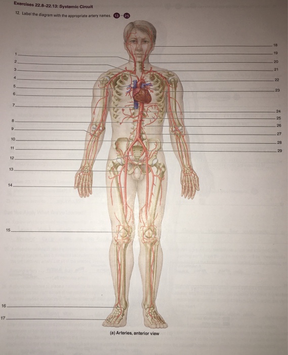
Solved: Label The Diagram With The Appropriate Artery Names. | Chegg.com from media.cheggcdn.com We hope this picture blood circulation principal veins and arteries diagram can help you study and research. These arteries arise in the neck, and ascend to the cranium. For more anatomy content please follow us and visit our website: There is a point at which the anterior and posterior arterial circuits of the brain unite or anastomose. The location of atherosclerotic lesions can be suggested by provocative stress testing (exercise or pharmacologic stress, and multiple evaluation modalities including ecg, echocardiography, and nuclear medicine). This allows blood to flow around the blocked artery to another artery nearby or to the same artery past the blockage, protecting the heart tissue from injury. Arteries and arterioles carry oxygenated blood _____ from the heart to the body. This area is known as the circle of willis.
Learn anatomy faster and remember everything you learn.
Defining the coronary artery anatomy is a critical step in any evaluation of ischemic heart disease and developing a treatment plan for your patient. This area is known as the circle of willis. Learn anatomy faster and remember everything you learn. It is a central communication that unites the internal carotid and vertebrobasilar systems. The cardiovascular system consists of the heart, blood vessels, and the approximately 5 liters of blood that the blood vessels transport. 5 out of 5 stars (293) 293 reviews $ 24.27. Anatomynote.com found human body artery diagram in detail from plenty of anatomical pictures on the internet. Learn the differences between an artery and a vein. We think this is the most useful anatomy picture that you need. Anatomynote.com found blood circulation principal veins and arteries diagram from plenty of anatomical pictures on the internet. Coronary arteries supply blood to the heart muscle. Arteries and veins are two of the body's main type of blood vessels. For more anatomy content please follow us and visit our website:
Arteries Diagram / Artery - Wikipedia / Artery, in human physiology, any of the vessels that, with one exception, carry oxygenated blood and nourishment from the heart to the tissues of the body.. There are any Arteries Diagram / Artery - Wikipedia / Artery, in human physiology, any of the vessels that, with one exception, carry oxygenated blood and nourishment from the heart to the tissues of the body. in here.